Diabetic Retinopothy
Diabetic Retinopathy and Diabetic Macular Edema
- Patients who have diabetes can develop diabetic retinopathy (DR). This occurs when blood sugar levels spike and remain high over time, damaging the blood vessels in the retina. DR is categorized into two main stages:
- Mild non-proliferative diabetic retinopathy (NPDR) is the earliest stage. In NPDR, tiny blood vessels may leak, causing the retina to swell. When this occurs in the macula, Macular Edema occurs. Macular Edema is a swelling of the macula thereby causing loss of vision.
- Proliferative diabetic retinopathy (PDR) is the more advanced stage of diabetic eye disease. In PDR, new blood vessels grow on the surface of the retina. This is also referred to as neovascularization. These blood vessels are very delicate and bleed easily and if remain untreated, the DR can cause scar tissue to develop on the retina, which then pulls on the retina, causing tractional retinal detachments.
- Diabetic retinopathy is the leading cause of vision-loss globally.1,2
- In adults (20-74), it is the leading cause of vision loss and is ranked the
fifth most common cause of preventable blindness.2,3 - Approximately 1/3 of diabetic patients (285 million worldwide) have signs of DR and of these, a further 1/3 is vision-threatening DR. This includes diabetic macular edema (DME).4
- A study in Wales revealed that patients with Type I Diabetes were at greater risk to development DR and PDR compared to those with Type II Diabetes, by more than 1/3. Of those screen, only 6% were found to have Type I Diabetes.5
- Lee R, Wong TY, Sabanayagam C. Epidemiology of diabetic retinopathy, diabetic macular edema and vision loss. Eye Vis (London) 2015;2:17.
- Cheung N, Mitchell P, Wong TY. Diabetic retinopathy. 2010;376(9735):124–36. doi: 10.1016/S0140-6736(09)62124-3. [PubMed] [Cross Ref]
- Bourne RR, Stevens GA, White RA, Smith JL, Flaxman SR, Price H, et al. Causes of vision loss worldwide, 1990–2010: a systematic analysis. Lancet Glob Health. 2013;1(6):e339–49. doi: 10.1016/S2214-109X(13)70113-X. [PubMed] [Cross Ref]
- Yau JW, Rogers SL, Kawasaki R, Lamoureux EL, Kowalski JW, Bek T, et al. Global prevalence and major risk factors of diabetic retinopathy. Diabetes Care.2012;35(3):556–64. doi: 10.2337/dc11-1909.[PMC free article] [PubMed] [Cross Ref]
- Thomas RL, Dunstan FD, Luzio SD, Chowdhury SR, North RV, Hale SL, et al. Prevalence of diabetic retinopathy within a national diabetic retinopathy screening service. Br J Ophthalmol. 2015;99(1):64–8. doi: 10.1136/bjophthalmol-2013-304017. [PubMed] [Cross Ref]
Treatment for Diabetic Macular Edema
Maculopexy© (Mx) or Combined Assisted Maculopexy (CAMP)
Hypothesis for the Mx effect
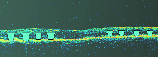
By using the laser as “glue” and gas for pressure, “Retinal Nails” form, into the retina at each laser spot, permanently restricting subsequent macular edema (thickening).

Image depicting “retinal nails” from laser applied, which essentially bonds the retinal layers together, forming a barrier to extravasted fluid from leaking blood vessels. This technique keeps the macula from absorbing or from “accepting” the hemorrhage from leaking blood vessels in the retina.
